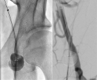
Figure 1
Bleeding complications at the access sites during catheter directed thrombolysis for acute limb ischaemia: Mini review
Elias Noory*, Tanja Böhme, Ulrich Beschorner and Thomas Zeller
Published: 03 March, 2021 | Volume 5 - Issue 1 | Pages: 001-003
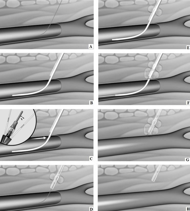
Figure 1:
Schematic representation of CaveoVasc® procedure. A. Guidewire in femoral artery. B. CaveoVasc® is applied after punction of the femoral artery and placement of the guide wire, thus in the beginning of the procedure. C. Removal of Locator, blood backflow indicates correct position. D. Infaltion of the Fixation Balloon – the inflated balloon secures the position of CaveoVasc® in the tissue. E. Placement of the sheath. F. Inflation of Pressure Balloon – Start of thrombolysis therapy via catheter perfusion. G. Removal of the sheath and catheter after the end of the thrombolysis procedure. H. Removal of CaveoVasc®.
Read Full Article HTML DOI: 10.29328/journal.avm.1001014 Cite this Article Read Full Article PDF
More Images
Similar Articles
-
Clinical characteristics in STEMI-like aortic dissection versus STEMI-like pulmonary embolismOscar MP Jolobe*. Clinical characteristics in STEMI-like aortic dissection versus STEMI-like pulmonary embolism. . 2020 doi: 10.29328/journal.avm.1001013; 4: 019-030
-
Bleeding complications at the access sites during catheter directed thrombolysis for acute limb ischaemia: Mini reviewElias Noory*,Tanja Böhme,Ulrich Beschorner,Thomas Zeller. Bleeding complications at the access sites during catheter directed thrombolysis for acute limb ischaemia: Mini review. . 2021 doi: 10.29328/journal.avm.1001014; 5: 001-003
Recently Viewed
-
Dairy cattle producers’ perception on Oestrus Synchronization and mass artificial insemination services in Waliso and Ilu Districts of South West Shoa Zone of Oromia, EthiopiaAbera Fekata*,Ulfina Galmessa,Lemma Fita,Chala Merera,Amanuel Bekuma. Dairy cattle producers’ perception on Oestrus Synchronization and mass artificial insemination services in Waliso and Ilu Districts of South West Shoa Zone of Oromia, Ethiopia. Insights Vet Sci. 2020: doi: 10.29328/journal.ivs.1001020; 4: 010-013
-
Exceptional cancer responders: A zone-to-goDaniel Gandia,Cecilia Suárez*. Exceptional cancer responders: A zone-to-go. Arch Cancer Sci Ther. 2023: doi: 10.29328/journal.acst.1001033; 7: 001-002
-
The prognostic value of p53 and WT1 expression in cancer: new molecular insights and epigenetics explanations lead to a new medical hypothesisAhed J Alkhatib* and Ilham Ahed Alkhatib. The prognostic value of p53 and WT1 expression in cancer: new molecular insights and epigenetics explanations lead to a new medical hypothesis. Arch Cancer Sci Ther. 2023: doi: 10.29328/journal.acst.1001034; 7: 003-009
-
Anticancer Activity of Genistin: A Short ReviewMd Mizanur Rahaman*, Md Iqbal Sikder, Muhammad Ali Khan and Muhammad Torequl Islam. Anticancer Activity of Genistin: A Short Review. Arch Cancer Sci Ther. 2023: doi: 10.29328/journal.acst.1001035; 7: 010-013
-
Knowledge, Attitude, and Practice of Healthcare Workers in Ekiti State, Nigeria on Prevention of Cervical CancerAde-Ojo Idowu Pius*, Okunola Temitope Omoladun, Olaogun Dominic Oluwole. Knowledge, Attitude, and Practice of Healthcare Workers in Ekiti State, Nigeria on Prevention of Cervical Cancer. Arch Cancer Sci Ther. 2024: doi: 10.29328/journal.acst.1001038; 8: 001-006
Most Viewed
-
Effects of dietary supplementation on progression to type 2 diabetes in subjects with prediabetes: a single center randomized double-blind placebo-controlled trialSathit Niramitmahapanya*,Preeyapat Chattieng,Tiersidh Nasomphan,Korbtham Sathirakul. Effects of dietary supplementation on progression to type 2 diabetes in subjects with prediabetes: a single center randomized double-blind placebo-controlled trial. Ann Clin Endocrinol Metabol. 2023 doi: 10.29328/journal.acem.1001026; 7: 00-007
-
Physical Performance in the Overweight/Obesity Children Evaluation and RehabilitationCristina Popescu, Mircea-Sebastian Șerbănescu, Gigi Calin*, Magdalena Rodica Trăistaru. Physical Performance in the Overweight/Obesity Children Evaluation and Rehabilitation. Ann Clin Endocrinol Metabol. 2024 doi: 10.29328/journal.acem.1001030; 8: 004-012
-
Hypercalcaemic Crisis Associated with Hyperthyroidism: A Rare and Challenging PresentationKarthik Baburaj*, Priya Thottiyil Nair, Abeed Hussain, Vimal MV. Hypercalcaemic Crisis Associated with Hyperthyroidism: A Rare and Challenging Presentation. Ann Clin Endocrinol Metabol. 2024 doi: 10.29328/journal.acem.1001029; 8: 001-003
-
Exceptional cancer responders: A zone-to-goDaniel Gandia,Cecilia Suárez*. Exceptional cancer responders: A zone-to-go. Arch Cancer Sci Ther. 2023 doi: 10.29328/journal.acst.1001033; 7: 001-002
-
The benefits of biochemical bone markersSek Aksaranugraha*. The benefits of biochemical bone markers. Int J Bone Marrow Res. 2020 doi: 10.29328/journal.ijbmr.1001013; 3: 027-031

If you are already a member of our network and need to keep track of any developments regarding a question you have already submitted, click "take me to my Query."


