Figure 2
Intravenous Leiomyomatosis of the Uterus with Intracardiac Extension
Tomas Reyes-del Castillo*, Minerva I Hernandez-Rejon, Jose L Ruiz-Pier, Mario Peñaloza-Guadarrama, Carlos E Merinos-Avila, Cristina Juarez-Cabrera, Pedro A del Valle-Maldonado, Sofia Ley-Tapia and Valentín Gonzalez-Flores
Published: 22 July, 2025 | Volume 9 - Issue 1 | Pages: 003-007

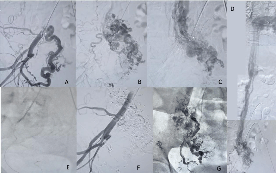
Figure 2:
A) Digital subtraction angiography of the right uterine artery showing a hypervascular tortuous tumor (B) with infiltration of the right ovarian vein (C) and its extension to the inferior vena cava and right auricle (F). (D) Selective cannulation of the ascending portion of the right uterine artery. E) Digital subtraction angiography showing complete occlusion of the tumor and the right uterine artery. F) Single-take radiograph showing hyperdense fluid emboli within the tumor.
Read Full Article HTML DOI: 10.29328/journal.avm.1001021 Cite this Article Read Full Article PDF
More Images
Similar Articles
-
The Impact of a Single Apheretic Procedure on Endothelial Function Assessed by Peripheral Arterial Tonometry and Endothelial Progenitor CellsGabriele Cioni*,Francesca Cesari,Rossella Marcucci,Anna Maria Gori,Lucia Mannini,Agatina Alessandrello Liotta,Elena Sticchi,Ilaria Romagnuolo,Rosanna Abbate,Giovanna D’Alessandri. The Impact of a Single Apheretic Procedure on Endothelial Function Assessed by Peripheral Arterial Tonometry and Endothelial Progenitor Cells. . 2017 doi: 10.29328/journal.hjvsm.1001001; 1: 001-007
-
Hepato-Pulmonary syndrome and Porto-Pulmonary Hypertension: Rare combination cause of Hypoxemia in patient with end-stage renal failure on Hemodialysis and hepatitis C Induced Decompensated CirrhosisAwad Magbri*,Mariam El-Magbri,Eussera El-Magbri. Hepato-Pulmonary syndrome and Porto-Pulmonary Hypertension: Rare combination cause of Hypoxemia in patient with end-stage renal failure on Hemodialysis and hepatitis C Induced Decompensated Cirrhosis. . 2017 doi: 10.29328/journal.avm.1001002; 1: 008-012
-
Management of Popliteal Artery aneurysms: Experience in our centerMichiels Thirsa,Vleeschauwer De Philippe*. Management of Popliteal Artery aneurysms: Experience in our center. . 2018 doi: 10.29328/journal.avm.1001003; 2: 001-009
-
Cystic adventitial disease of the external iliac artery with disabling claudication: A case report and short reviewNalaka Gunawansa*. Cystic adventitial disease of the external iliac artery with disabling claudication: A case report and short review. . 2018 doi: 10.29328/journal.avm.1001004; 2: 010-013
-
Anesthetic considerations for endovascular repair of ruptured abdominal aortic aneurysmsKH Kevin Luk*,Koichiro Nandate. Anesthetic considerations for endovascular repair of ruptured abdominal aortic aneurysms. . 2018 doi: 10.29328/journal.avm.1001005; 2: 014-019
-
Severe aorto-iliac occlusive disease: Options beyond standard aorto-bifemoral bypassKonstantinos Filis*,Constantinos Zarmakoupis,Fragiska Sigala,George Galyfos . Severe aorto-iliac occlusive disease: Options beyond standard aorto-bifemoral bypass. . 2018 doi: 10.29328/journal.avm.1001006; 2: 020-024
-
Navigation in the land of ScarcityAwad Magbri*,Mariam El-Magbri. Navigation in the land of Scarcity. . 2018 doi: 10.29328/journal.avm.1001007; 2: 025-026
-
Transcatheter embolization of congenital vascular malformations, single center experienceMohammed Habib*,Majed Alshounat. Transcatheter embolization of congenital vascular malformations, single center experience. . 2019 doi: 10.29328/journal.avm.1001008; 3: 001-006
-
A fatal portal vein thrombosis: A case reportM Kechida*,W Ben Yahia,W Mnari. A fatal portal vein thrombosis: A case report. . 2019 doi: 10.29328/journal.avm.1001009; 3: 007-008
-
PR and QT intervals short on the same electrocardiogramFrancisco R Breijo-Márquez*. PR and QT intervals short on the same electrocardiogram. . 2020 doi: 10.29328/journal.avm.1001010; 4: 001-004
Recently Viewed
-
Unveiling Disparities in WHO Grade II Glioma Care among Physicians in Middle East and North African (MENA) Countries: A Multidisciplinary SurveyFatimah M Kaabi,Layth Mula-Hussain*,Shakir Al-Shakir,Sultan Alsaiari,Leonidas Chelis,Renda AlHabib,Sara Owaidah,Renad Subaie,Marwah M Abdulkader,Ibrahim Alotain. Unveiling Disparities in WHO Grade II Glioma Care among Physicians in Middle East and North African (MENA) Countries: A Multidisciplinary Survey. Arch Cancer Sci Ther. 2026: doi: 10.29328/journal.acst.1001048; 10: 001-005
-
Maximizing the Potential of Ketogenic Dieting as a Potent, Safe, Easy-to-Apply and Cost-Effective Anti-Cancer TherapySimeon Ikechukwu Egba*,Daniel Chigbo. Maximizing the Potential of Ketogenic Dieting as a Potent, Safe, Easy-to-Apply and Cost-Effective Anti-Cancer Therapy. Arch Cancer Sci Ther. 2025: doi: 10.29328/journal.acst.1001047; 9: 001-005
-
Analysis and Control of a Glucose-insulin Dynamic ModelLakshmi N Sridhar*. Analysis and Control of a Glucose-insulin Dynamic Model. Ann Clin Endocrinol Metabol. 2026: doi: 10.29328/journal.acem.1001033; 10: 010-016
-
Transumbilical Single-incision Hiatal Hernia Repair and Nissen Fundoplication in situs Inversus Totalis: A Rare Case ReportQing Cao,Chen Kang,Kang Gu,Yin Peng,Yang Lv,Xu-Zhong Ding,Peng Li*. Transumbilical Single-incision Hiatal Hernia Repair and Nissen Fundoplication in situs Inversus Totalis: A Rare Case Report. Adv Treat ENT Disord. 2026: doi: 10.29328/journal.ated.1001017; 10: 001-003
-
NAD⁺ Biology in Ageing and Chronic Disease: Mechanisms and Evidence across Skin, Fertility, Osteoarthritis, Hearing and Vision Loss, Gut Health, Cardiovascular–Hepatic Metabolism, Neurological Disorders, and MuscleRizwan Uppal,Umar Saeed*,Muhammad Rehan Uppal. NAD⁺ Biology in Ageing and Chronic Disease: Mechanisms and Evidence across Skin, Fertility, Osteoarthritis, Hearing and Vision Loss, Gut Health, Cardiovascular–Hepatic Metabolism, Neurological Disorders, and Muscle. Ann Clin Endocrinol Metabol. 2026: doi: 10.29328/journal.acem.1001032; 10: 001-009
Most Viewed
-
Impact of Latex Sensitization on Asthma and Rhinitis Progression: A Study at Abidjan-Cocody University Hospital - Côte d’Ivoire (Progression of Asthma and Rhinitis related to Latex Sensitization)Dasse Sery Romuald*, KL Siransy, N Koffi, RO Yeboah, EK Nguessan, HA Adou, VP Goran-Kouacou, AU Assi, JY Seri, S Moussa, D Oura, CL Memel, H Koya, E Atoukoula. Impact of Latex Sensitization on Asthma and Rhinitis Progression: A Study at Abidjan-Cocody University Hospital - Côte d’Ivoire (Progression of Asthma and Rhinitis related to Latex Sensitization). Arch Asthma Allergy Immunol. 2024 doi: 10.29328/journal.aaai.1001035; 8: 007-012
-
Causal Link between Human Blood Metabolites and Asthma: An Investigation Using Mendelian RandomizationYong-Qing Zhu, Xiao-Yan Meng, Jing-Hua Yang*. Causal Link between Human Blood Metabolites and Asthma: An Investigation Using Mendelian Randomization. Arch Asthma Allergy Immunol. 2023 doi: 10.29328/journal.aaai.1001032; 7: 012-022
-
An algorithm to safely manage oral food challenge in an office-based setting for children with multiple food allergiesNathalie Cottel,Aïcha Dieme,Véronique Orcel,Yannick Chantran,Mélisande Bourgoin-Heck,Jocelyne Just. An algorithm to safely manage oral food challenge in an office-based setting for children with multiple food allergies. Arch Asthma Allergy Immunol. 2021 doi: 10.29328/journal.aaai.1001027; 5: 030-037
-
Snow white: an allergic girl?Oreste Vittore Brenna*. Snow white: an allergic girl?. Arch Asthma Allergy Immunol. 2022 doi: 10.29328/journal.aaai.1001029; 6: 001-002
-
Cytokine intoxication as a model of cell apoptosis and predict of schizophrenia - like affective disordersElena Viktorovna Drozdova*. Cytokine intoxication as a model of cell apoptosis and predict of schizophrenia - like affective disorders. Arch Asthma Allergy Immunol. 2021 doi: 10.29328/journal.aaai.1001028; 5: 038-040

If you are already a member of our network and need to keep track of any developments regarding a question you have already submitted, click "take me to my Query."


